ドキュメンテーション
Therapeutic strategies can only be efficiently discussed if the diagnoses are objectively documented. The fact that this is now a matter of routine is due to KARL STORZ. Scientific progress would be unthinkable without a rapid, comprehensive flow of information on diagnostic findings.
The KARL STORZ enterprise has developed technologies for making diagnostic results immediately accessible to all parties involved in the treatment process.
KARL STORZ pioneered image-supported documentation of veterinary medical diagnoses initially with the aid of flash devices for 35 mm cameras and, in later years, with high powered video systems for use within the sterile area, and gained valuable experience with these systems.
Digital technology has already replaced many conventional documentation media. With the development of the integrated operating room, digital data archiving and global communication from the operating room, KARL STORZ is now pioneering applications which tomorrow will be a matter of routine.
The range of KARL STORZ products comprises state-of-the-art fully digital 3-Chip-Cameras along with mobile ALL-in-ONE solutions for the medical practice, together with the necessary peripheral devices such as monitors, video recorders and video printers. For the all-important field of illumination, KARL STORZ offers a range of unparalleled diversity that covers the entire spectrum of possible applications – from the high-power xenon light source to the mobile battery light source.
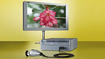
IMAGE1 S™ 4U – mORe than a camera
The IMAGE1 S™ 4U camera system allows the operating surgeon to make optimal use of the benefits offered by 4K technology. A notable feature is the image quality: High image brightness, impressive colors, greater richness of detail and a significantly improved depth effect characterize this system. Thanks to the system’s modularity, 4U components can be easily integrated into the existing IMAGE1 S™ camera platform. Consequently, the system is still compatible with existing technologies (e.g., rigid, flexible, fluorescence and 3D endoscopy) and can be adapted to meet individual customer needs.
- IMAGE1 S™ 4U impresses with outstanding, razor-sharp images
- Excellent image brightness
- First-rate color rendition
- Greater richness of detail
- Three innovative visualization technologies for tissue differentiation:
- CLARA: Homogeneous illumination
- CHROMA: Contrast enhancement
- SPECTRA*: Spectral color shift and switch
- Easy integration into the IMAGE1 S™ camera platform
* not for sale in the U.S.
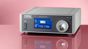
CO2mbi® LED Cold Light Fountain
- Two-in-one solution combines a LED light source and air/CO2 insufflator in one unit
- Long LED module service life provides significant cost reductions
- Manually adjustable light intensity
- Optimal light yield with high-efficiency LED
- CO2 insufflation for greater patient comfort
- Choice of three insufflation modes: 100% CO2 mode, regular room air,
- CO2 /air mode
- Integrated pump with two power levels for rapid insufflation
- Display with interactive icons
- Touch screen can also be controlled with wet gloves
VITOM® 25
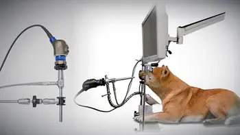
The VITOM® 25 exoscope offers a revolutionary new way of displaying open surgical procedures in an ergonomic and high quality manner. Unlike a traditional endoscope, the VITOM® 25 is an “exoscope” which is placed at a distance of 25 to 75 cm from the surgical site, held securely in place by a holding device, giving the surgeon ample workspace.
The magnified image produced by the VITOM® 25 is viewed on a video monitor. This enables the surgeon and support staff to work together comfortably, significantly reducing surgeon fatigue, while the magnification of structures improves surgical precision and accuracy of diagnosis.
This system is ideal for teaching, as is allows for easy, magnified viewing of the surgery, which can also be transmitted to remote locations, without disruption of the procedure.
The ability to capture and archive images and videos of surgical procedures ensures that a detailed and accurate record of the diagnosis and treatment performed is recorded, while creating a valuable tool for training, education, re-examination at a later time, and sharing with clients and colleagues.
The VITOM® 25 works with any existing KARL STORZ video system.


