Secondary Prevention of Cervical Cancer
Border – Major Change
A sharp bordered lesion is decribed as consistent with major change.
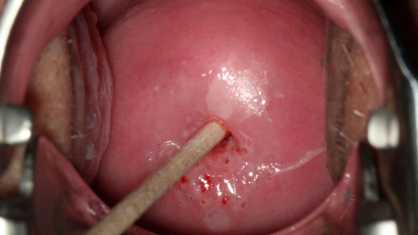
The sharp colposcopic delineation of a lesion is associated with the presence of high-grade SIL. Colposcopic findings in a 26-year-old gravida 0 who had not been vaccinated against HPV. Her cervical smear was reported as class IIID1. High risk HPV DNA (not type 16 or 18) was detected. Colposcopy shows a sharply marginated acetowhite lesion at 12–1 o’clock in an atypical T-zone type 2, which is classified as grade 2 (major change) abnormal colposcopic finding.
Squamous epithelium and SIL are clearly separated in CIN 3 .

Histopathology of tissue sampled by loop biopsy shows atypical squamous epithelium in which expression of the immune markers Ki67 and p16 extends into the highest cell layer. This is also apparent in the involvement of an endocervical crypt (gland) at the bottom of the image. Note the sharp delineation from normal squamous epithelium at the left edge of the image. The histopathologic diagnosis is grade 3 cervical intraepithelial neoplasia (CIN 3).