Secondary Prevention of Cervical Cancer
Metaplasia – A Physiological Process
Metaplasia – A Physiological Process
Indirect metaplasia replaces columnar epithelium by squamous epithelium.
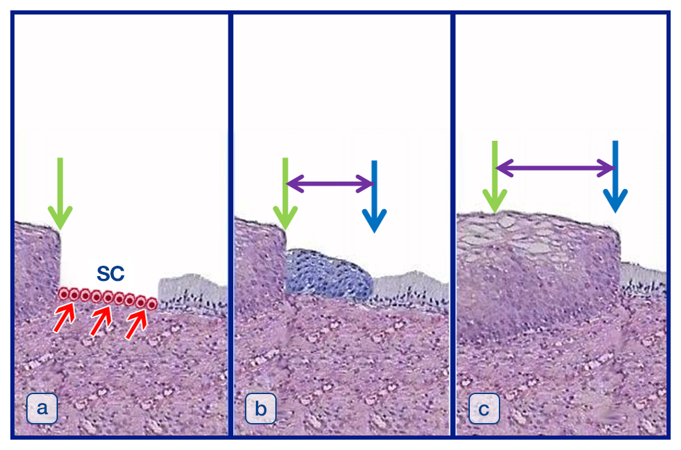
Histology shows the squamous epithelium on the left side bordering the columnar epithelium on the right side. The green arrow indicates the congenital junction which is identical to the squamocolumnar junction before puberty. In (a), red arrows point to squamocolumnar junction cells (SC) which have the biologic potential of stem cells. In (b), the squamocolumnar junction cells have undergone metaplasia and metaplastic epithelium is present. Thus, the congenital junction is still visible next to the metaplastic epithelium, indicated by the green arrow. Now, the squamocolumnar junction has shifted and is called adult or functional junction, indicated by the blue arrow. The purple arrows indicate the extent of the transformation zone (T-zone) which lies in between the congenital and the adult or functional junction. In (c), metaplastic epithelium has been transformed to squamous epithelium. Thus, the left portion of the squamous epithelium is original squamous epithelium, the right portion of the squamous epithelium is secondary squamous epithelium since it originated from metaplasia. This whole area is the T-zone, indicated by the purple arrrows. The congenital junction indicated by the green arrow is no longer distinguishable in terms of histopathology. The squamocolumnar junction is identical to the adult or functional junction indicated by the blue arrow and may shift more to the right with age.