Secondary Prevention of Cervical Cancer
Evaluating a Smear
Using light microscopy basal cells, parabasal cells, intermediate cells and/or superficial cells can be identified. In addition columnar cells and in the presence of metaplasia metaplastic cells can be seen. In the presence of precancer dyskaryotic cells are present.
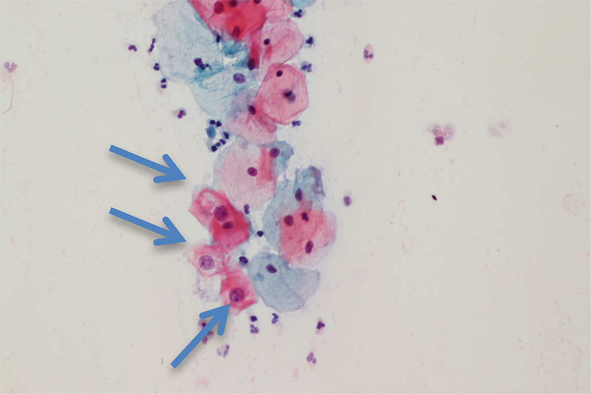
The cytophotogram shows three dyskaryotic cells with an halo in the cytoplasm, so-called koilocytes, which are marked by arrows can be seen. The nuclei of these koilocytes contain papillomavirus particles. The ratio between the nucleus and cytoplasm is increased in the koilocyte compared to the normal cell. Therefore these cells are mature dyskaryotic cells. This Pap smear is suggestive of CIN 1 or low-grade SIL. According to the Munich III classification, this smear is classified as class IIID1.
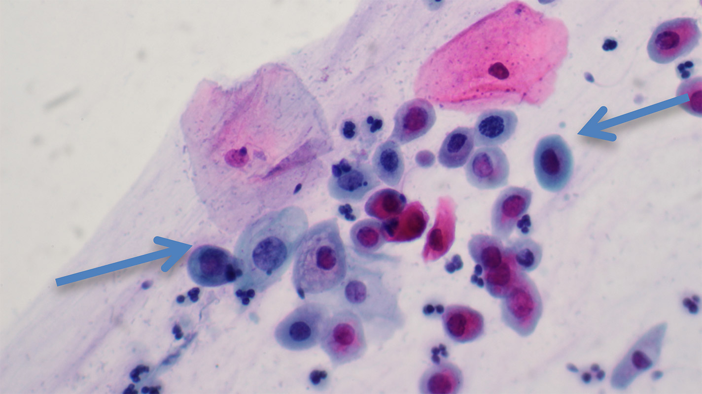
This cytophotogram shows two normal eosinophilic red-stained superficial cells in the upper part. Below there is a syncytium of severe dyskaryotic cells (marked by arrows) with a highly distorted ratio between nucleus and cytoplasm marked by arrows. The nuclei are hyperchromatic and anisokaryotic. This Pap smear is suggestive of CIN 3 or high-grade SIL. According to the Munich III classification, this smear is classified as IVa-p.